How to Read a Breast Cancer Pathology Report: A Practical Guide for Clinicians
A breast cancer pathology report is far more than a document confirming a diagnosis of malignancy. It is the definitive clinical map guiding the entire multidisciplinary team—from surgical choices and radiation planning to systemic treatment and genetic counseling.
Yet, under the pressure of busy clinics, pathology reports are often read selectively: a quick glance at the diagnosis, size, nodal status, and core receptors (ER, PR, HER2), and treatment begins. By reading selectively, clinicians can easily overlook critical findings that significantly impact patient prognosis and care.
This guide provides a structured framework for oncologists and general practitioners (GPs) to systematically interpret a breast cancer pathology report, understand the "why" behind key reporting protocols, and spot the subtle features that dictate advanced molecular or germline testing.
The Four Levels of the Diagnostic Pyramid
To avoid both under-testing and unnecessary over-testing, think of breast cancer tissue analysis as a series of progressively deeper levels of investigation. Each level answers a distinct clinical question:
- Level 1: Histopathology – What does the tumor look like? This evaluates physical tissue structure, architectural morphology, and physical margins.
- Level 2: Immunohistochemistry (IHC) & Predictive Biomarkers – What proteins does the tumor express? This defines the basic biological behavior and surface receptors.
- Level 3: Tissue-Based Molecular & Genomic Testing – What is the tumor's genetic signature? This estimates recurrence risk and predicts chemotherapy benefit.
- Level 4: Germline Genetic Testing – Did the patient inherit a genetic mutation? This uses normal cells (blood/saliva) to assess systemic cancer risks for the patient and their family.
Level 1: Core Histopathology & The CAP Protocol
Histopathology is the microscopic evaluation of tissue sections stained with hematoxylin and eosin (H&E stain). To prevent data fragmentation and ensure complete clarity across institutions, modern pathology departments rely on standardized reporting templates.
The CAP Protocol vs. ASCO/CAP Guidelines
It is important to keep these two separate:
- CAP Cancer Protocols: Standardize what data points must be physically documented in the synoptic checklist (e.g., margins, tumor type, grading metrics) to prevent data gaps.
- ASCO/CAP Guidelines: Provide the exact scientific criteria for how specific predictive biomarkers (like HER2 or ER) are analyzed and assigned a clinical status.
Core Tumor Characteristics to Check:
- Histological Type: While most cases are Invasive Breast Carcinoma of No Special Type (IBC-NST)—formerly called invasive ductal carcinoma—look out for special types like invasive lobular carcinoma (which frequently presents with multifocal, hard-to-image growth), mucinous, tubular, or aggressive metaplastic carcinomas.
- Nottingham Histologic Score (Tumor Grade): Measures tubule formation, nuclear pleomorphism (cell shape variation), and mitotic activity to classify the tumor from Grade 1 to Grade 3. It is vital to note that tumor grade reflects morphological aggressiveness and mitotic activity under the microscope, which is independent of the anatomical stage of the disease.
- Invasive Component Size: Staging criteria require measuring only the largest contiguous focus of invasive carcinoma. Do not mistake the total specimen size or adjacent ductal carcinoma in situ (DCIS) for the true invasive measurement.
- Lymphovascular Invasion (LVI): The presence of tumor cells within endothelial-lined lymphatic or vascular channels. While LVI does not change the anatomical TNM stage, it is an independent adverse prognostic factor that influences locoregional management decisions.
- Surgical Margins: The distance from malignant cells to the inked outer edge of the specimen. Margins less than 2mm are considered close or involved, requiring direct correlation with the surgical approach (breast-conserving surgery vs. mastectomy) and consensus guidelines.
Level 2: The Lymph Node Report—Beyond "Positive" or "Negative"
Axillary lymph node status remains a foundational prognostic pillar. A complete lymph node analysis requires a deep look into the structural burden of the regional disease:
-
Size Thresholds of Metastatic Deposits:
- Macrometastases (>2.0 mm): Clearly defined, large deposits driving standard nodal staging.
- Micrometastases (0.2 mm to 2.0mm): Smaller clusters that still represent low-volume nodal disease.
- Isolated Tumor Cells (ITCs, <0.2mm): Tiny single cells or clusters that do not count as true structural metastases for conventional staging but are carefully noted.
- Extranodal Extension (ENE): This occurs when tumor cells breach the fibrous lymph node capsule into neighboring adipose tissue. ENE is an adverse feature strongly associated with high local recurrence rates, often prompting a transition from a simple sentinel lymph node biopsy to a complete axillary dissection, along with modified regional radiation fields.
- Post-Neoadjuvant Treatment Response: Following neoadjuvant systemic therapy, look for documented signs of regression (like residual fibrosis or histiocytic responses). Even if no viable tumor remains, these signs provide evidence of previous nodal disease burden.
Level 3: Immunohistochemistry (IHC) & Predictive Biomarkers
IHC uses targeted antibodies to identify specific proteins on or inside the tumor cells. Every primary breast path report requires an internal and external control verification for four standard markers:
| Biomarker | Clinical Role | Critical Cut-Offs & Interpretation |
|---|---|---|
| Estrogen Receptor (ER) | Predicts responsiveness to endocrine therapies (e.g., tamoxifen, aromatase inhibitors). | > 1% nuclear staining is positive. Note if it falls in the 1-10% "ER-low-positive" category, as these often behave like hormone-receptor-negative tumors. |
| Progesterone Receptor (PR) | Reflects a functional, intact estrogen receptor pathway. | Evaluated via staining percentage; helps distinguish Luminal A from Luminal B subtypes. |
| HER2 | Detects human epidermal growth factor receptor 2 protein overexpression. | 0 or 1+: Negative 2+: Equivocal (Triggers automatic reflex testing) 3+: Positive |
| Ki-67 Proliferation Index | Measures the percentage of cells actively dividing. | Interpreted alongside grade and stage. Scores >20% indicate highly proliferative, aggressive disease. |
When to Order Tissue-Based Molecular & Genomic Assays
If a tumor is HER2 2+ (Equivocal), reflex testing via Fluorescence In Situ Hybridization (FISH) is required to check for actual gene amplification.
For patients with ER/PR-positive, HER2-negative, lymph-node-negative early-stage breast cancer, routine clinical features may not clearly show whether chemotherapy adds any survival benefit. In these cases, multigene expression profiling is highly valuable:
- Oncotype DX: Evaluates a 21-gene signature to provide a Recurrence Score (0-100). This test serves both a prognostic role (predicting 10-year distant recurrence risk) and a predictive role (quantifying exactly who will benefit from adjuvant chemotherapy).
- MammaPrint: A 70-gene expression profile that categorizes patients strictly into high vs. low genomic risk groups.
- PAM50 (ProSigna): Analyzes 50 genes to identify the biological intrinsic subtype and provides a definitive Risk of Recurrence (ROR) score.
- EndoPredict & Breast Cancer Index (BCI): Excellent genomic toolkits for evaluating late recurrence risks and determining the benefit of extended endocrine therapy beyond 5 years.
Level 4: Somatic vs. Germline Testing—When to Look for BRCA
It is critical to distinguish between somatic testing (evaluating genetic changes unique to the tumor cells to find actionable drug targets like PIK3CA or ESR1) and germline testing (evaluating inherited mutations present in every cell of the patient's body).
Triggers for Germline Genetic Testing
A patient's normal blood or saliva should be tested for inherited variants if they meet any of the following clinical criteria:
- Diagnosed at age 50 or younger.
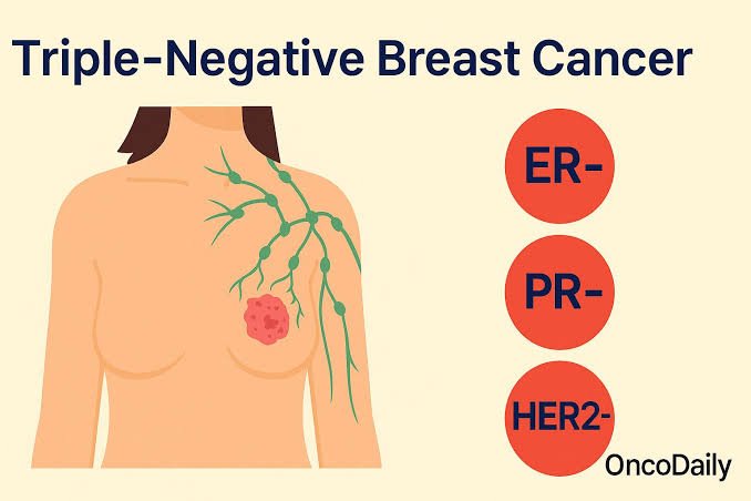
- Diagnosed with Triple-Negative Breast Cancer (TNBC) at any age.
- Presents with multiple primary breast cancers (synchronous or metachronous).
- Has a personal or strong family history of male breast cancer, ovarian, pancreatic, or metastatic prostate cancer.
Why an HBOC Multigene Panel Beats Standalone BRCA Testing
Historically, genetic testing was limited to checking BRCA1 and BRCA2. Modern standard of care requires a comprehensive Hereditary Breast and Ovarian Cancer (HBOC) multigene panel.
The Clinical Advantage: Hereditary breast cancer is highly heterogeneous. An HBOC panel checks BRCA1/2 alongside other high- and moderate-penetrance genes like PALB2, CHEK2, ATM, TP53, and PTEN.
Note on Variants of Uncertain Significance (VUS): Broader panels increase the chance of finding a VUS. A VUS indicates a regular genetic variation of unknown clinical impact and should never be used to justify major surgical interventions like prophylactic bilateral mastectomies.
The 12-Point Clinical Pathology Checklist
When reviewing a new breast cancer pathology report in your clinic, run through this quick, systematic checklist before finalizing a treatment strategy:
- Diagnosis: Is it invasive, in situ (DCIS), or an alternate lesion?
- Histological Type: Is it a standard type (IBC-NST) or a variant like lobular or metaplastic?
- Nottingham Grade: What is the specific grade assigned (Grade 1, 2, or 3)?
- Invasive Tumor Size: What is the exact size of the true invasive component in millimeters?
- Lymphovascular Invasion (LVI): Is it clearly documented as present or absent?
- Surgical Margins: What is the precise distance to the closest inked margin for both invasive and DCIS components?
- Nodal Count: How many total nodes and sentinel nodes were retrieved and analyzed?
- Nodal Deposit Size: Are the nodal deposits classified as macrometastases, micrometastases, or ITCs?
- Extranodal Extension (ENE): Is there any evidence of tumor spread beyond the node capsule?
- Hormone Status (ER/PR): What are the exact staining percentages? Does the patient fall into the ER-low-positive range?
- HER2 Value: Is the IHC score 0, 1+, 2+, or 3+? If 2+, has reflex FISH testing been completed?
- Ancillary Assays: Does this patient meet the criteria for a tumor genomic assay (e.g., Oncotype DX) or an inherited germline HBOC multigene panel?
Ultimately, a pathology report is not an administrative hurdle—it is a detailed biological map of the disease. Developing a systematic approach to reading ensures that no prognostic marker is missed and every treatment plan is tailored precisely to the patient's unique tumor biology.


How to Read a Breast Cancer Pathology Report: A Practical Guide for Clinicians
Is It a Mistake to Operate on Triple-Negative Breast Cancer Upfront?
Best Cancer Surgeon in Mysore – How to find one; 7 points Patients Should Know Before Choosing a Surgeon

10 Powerful Cancer prevention tips – A Doctor’s Guide to a Healthier Life